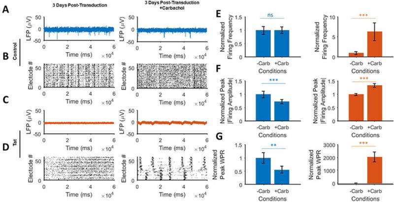
Figure 6. Activating the acetylcholine receptors in the absence and presence of Tat.

A. Extracellular action potential recordings before (left) and after (right) application of the AChR receptors agonist, carbachol (carbamylcholine) in the control (Ad-Null) group 3 days post-transduction. Carbachol partially increases the spontaneous firing while reducing the firing amplitude. B. Raster plots of the detected spikes show increased asynchronous activity (right) in the control group compared to the baseline activity (left). C. Extracellular action potential recordings before (left) and after (right) application of carbachol in the Tat expressing group 3 days post-transduction. Application of carbachol gives rise to increased firing activity (left) as compared to the pre-carbachol condition (left). D. Detected spikes raster plots of the Tat expressing neurons show the generation of regularly spaced spike trains (right) as opposed to no spontaneous firing (left) before carbachol application. E. Carbachol application shows a negligible effect on the control (left) compared to the Tat expressing groups (right, increased amplitude). F. There is a decrease in the spike frequencies for the control group (left) and increase in the Tat expressing group (right). G. Normalized peak WPRs derived from the electrodes data analyses show a significant increase in Tat expressing neurons (right), with a relative decrease in the control group (right). Quantified data show mean ± StDev as determined by student t-test, n.s. not significant, **= p<0.01, ***= p<0.001.
