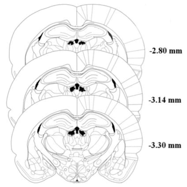
Figure 5.
Histological confirmation of bilateral cannula placements within the lateral habenula. Shaded regions on either side of the third ventricle represent areas in which intracranial cannula tips were successfully localized to the target region. Figure adapted from [74]. Numbers represent the distance of each coronal slice from bregma.

