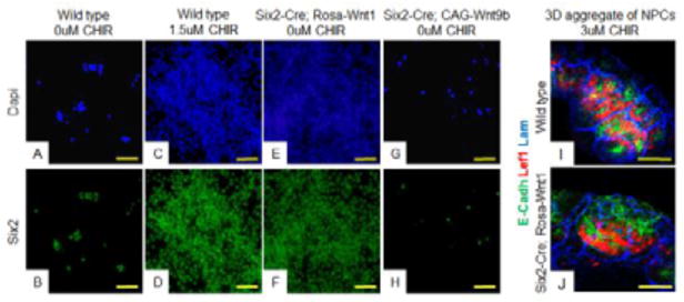
Figure 5. Distinct beta-catenin activity levels induce distinct cell fates on isolated, pure NPC cultures.
Wildtype (A–D, I), Six2-Cre;Rosa-Wnt1 (E–F, J,) or Six2-Cre;CAG-Wnt9b (G–H,) purified NPCs were cultured in media with 1.5uM CHIR (C–D,), 0uM CHIR (A–B, E–H,) or 3uM CHIR (I–J) and stained with antibodies to the NPC marker Six2 (green) (B–H), differentiation marker Lef1 (red in I,J), epithelial tubule marker E-cadherin (green in I,J), or basement membrane marker Laminin (blue in I,J). In I and J, cells were aggregated and grown at the air media interface for two days. All images are counterstained with DAPI (blue). Scale bars are 100uM.

