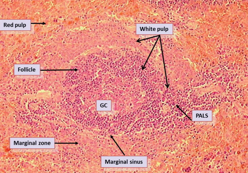
FIG. 1.
Histological structural organization of the rat spleen. The spleen is divided into red pulp (RP) and white pulp (WP) regions. The WP are further divided into B and T lymphocyte regions including the follicles, marginal zone and periarteriole lymphatic sheath (PALS). Visible in the WP are a secondary follicle i.e., a follicle containing a germinal center. The marginal zone (MZ) forms an interface between the RP and the WP. The MZ is further separated from the follicle and PALS by the marginal sinus.

