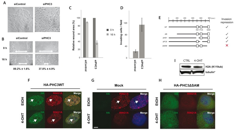
Figure 16. PHC3 depletion alters RING1A localization, cellular motility and invasion.
Cells treated with either non-targeting siRNA (siControl), or siRNA targeting PHC3, were analysed by light microscopy for ( A) morphological changes, ( B, C) wound healing assays, and ( D) transwell Matrigel invasion assays. Error bars indicate standard error between three biological replicates. E) A summary of data comparing the ability of full length PHC3 and PHC3 truncation mutants to repress the invasion phenotype induced by v-Src activation. ( F, G, H) Cells were transduced with lentivirus encoding for either ( F) HA-PHC3WT (full length, wild-type), empty ( G), or ( H) HA-PHC3ΔSAM (SAM domain deletion mutant). Cells were then treated either with ethanol (control), or 4-OHT, and immunostained for HA tag (green) and RING1A (red). Arrows indicate RING1A nuclear foci. Representative images of three replicates. Scale bar: 5 µm. ( I) Immunoblot analysis of H2AK119ub -/+ Src activation. Note that the loading control shown is identical to Figure 15E.

