Figure 7.

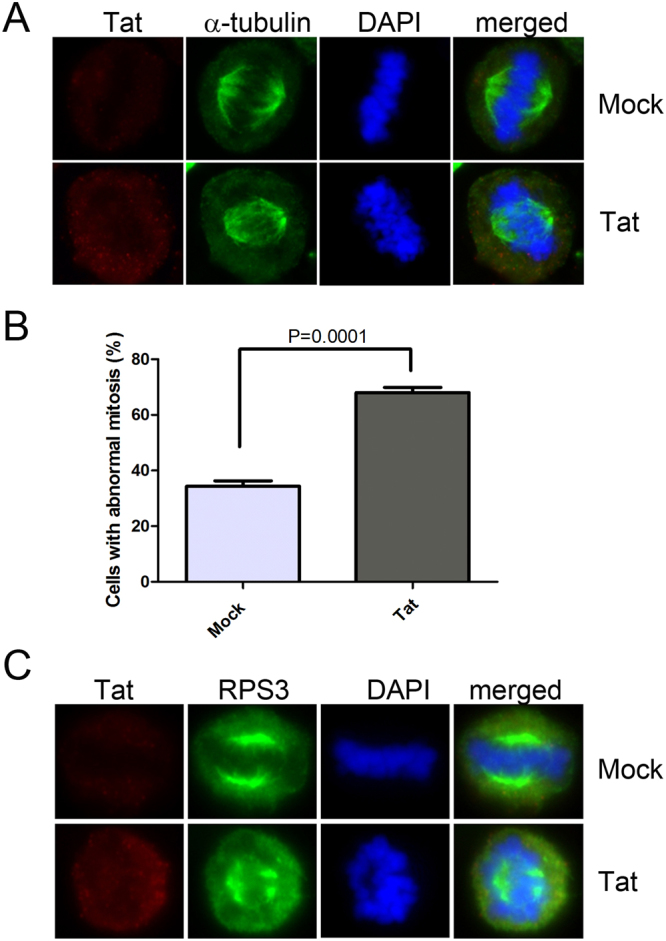
HIV-1 Tat disturbs mitotic spindle formation in Jurkat cells. (A) A representative figure of the mitotic spindle defect in Lenti-Tat-transduced Jurkat cells. Jurkat cells were transduced with the control virus or Lenti-Tat and immunostained with antibodies against Tat and α-tubulin 3 days after infection. Control, n = 189; Tat, n = 220 from three independent experiments. (B) Mitotic defects in Jurkat cells. The percentage of cells showing mitotic spindle formation defects and misaligned chromosomes is plotted. Data are presented as mean ± SEM from three independent experiments. Statistical analysis was performed by an unpaired t test. Control, n = 189; Tat, n = 220; P < 0.0001. (C) Localization of RPS3 in the mitotic spindle in the presence of Tat. Lenti-Tat-infected Jurkat cells were immunostained with antibodies against Tat and RPS3 at 3 days after infection. RPS3 was displaced from the mitotic spindle pole, and an aberrant spindle was formed. Control, n = 44; Tat, n = 132. Experiments were repeated two times.
