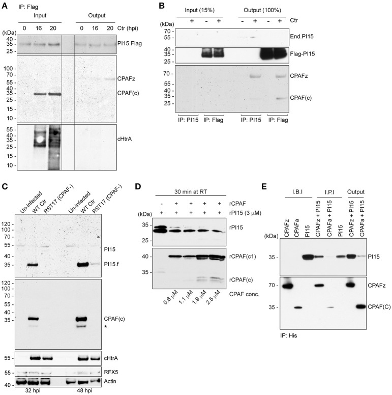
Figure 4.
PI15 interacts with the CPAF zymogen. (A) PI15 binds to the CPAF zymogen. Co-immunoprecipitation (Co-IP) experiments were carried out using HeLa cells that were transiently transfected with a PI15-Flag constructs. Cells were infected with C. trachomatis for different time intervals. Total cellular lysates were prepared and used for Co-IP using Flag beads. (B) PI15 interacts with CPAF. Co-IP experiments were carried out using T-REx-293 cells that inducibly express PI15-Flag. Cells were induced with 0.1 μg/ml of AHT for 48 h and then infected with C. trachomatis for another 24 h. Total cellular lysates were prepared and used for Co-IP using either commercial Flag beads or agarose beads with covalently cross-linked PI15 antibodies. (C) Degradation of PI15 depends on CPAF. HeLa cells were infected with Chlamydia wild type or the Chlamydia CPAF mutant RST17 (CPAF−), for 32 or 48 h, respectively. PI15 as well as CPAF expression was analyzed by immunoblotting. Post lysis degradation of substrates by CPAF was monitored by testing RFX5 expression. *, PI15 bands from the previous blot. (D) CPAF cleaves PI15 in vitro. Recombinant PI15 was incubated in the presence of increasing concentrations of recombinant CPAF at room temperature for 30 min and were analyzed by immunoblotting. (E) CPAF interacts with PI15 in vitro. Co-IP was carried out using an inactive full-length recombinant CPAF or active CPAF (both His-tagged) and recombinant PI15 (without any tag). I.B.I., Input samples before incubation; I.P.I., Input after 2 h incubation at room temperature; CPAFz, CPAF zymogen; CPAFa, CPAF active; CPAF(c), c-terminal fragment of active CPAF; CPAF(c1), intermediate c-terminal fragment of active CPAF; End.PI15, endogenous PI15.

