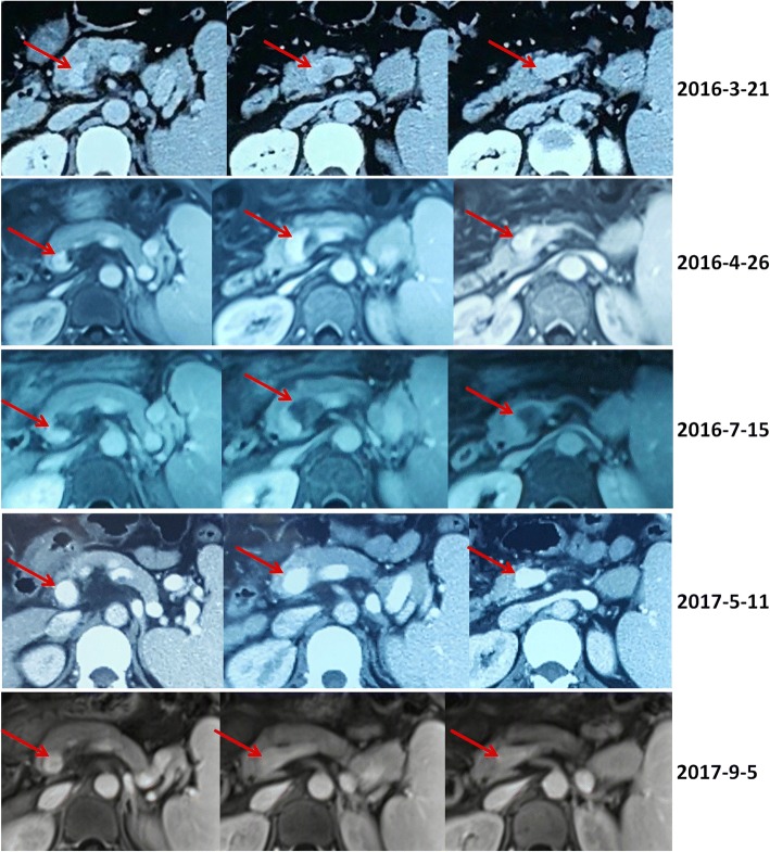
Fig. 2.
Contrast-enhanced computed tomography and magnetic resonance scans in a patient with transient PVT. Contrast-enhanced computed tomography and magnetic resonance scans performed in 2016 demonstrated a partial thrombosis within the confluence of portal vein and splenic vein (red arrows). Contrast-enhanced computed tomography and magnetic resonance scans performed in 2017 demonstrated that the confluence of portal vein and splenic vein was patent (red arrows)

