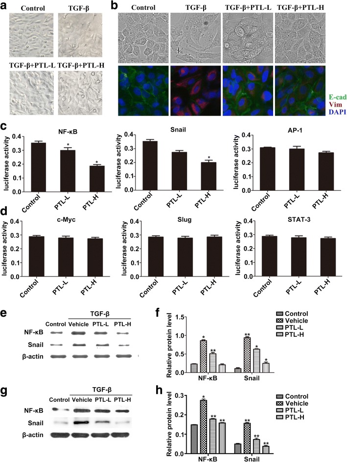
Fig. 2.
PTL inhibits TGF-β1-induced EMT through inhibiting NF-κB/Snail expression in lung epithelial cells. a Typical images of A549 cells in the Control group, TGF-β group, PTL-L + TGF-β group, and PTL-H + TGF-β group under an optical microscope. b Typical images of primary lung epithelial cells in the Control group, TGF-β group, PTL-L + TGF-β group, and PTL-H + TGF-β group under an optical microscope and immunofuorescence staining of E-Cadherin (green) and Vimentin (red) was performed. The nucleus was staining with DAPI. c, d Expression levels of NF-κB, Snail, AP-1, c-Myc, Slug and Stat-3 were assessed using dual-luciferase assay. e-h After TGF-β/PTL treatment, NF-κB and Snail were evaluated using Western blot analysis. β-actin was used as a loading control. Data are presented as means of three experiments; error bars represent standard deviation, *P < 0.05, **P < 0.01

