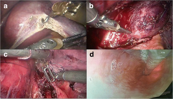
Fig. 3.
Robot-assisted enucleation of a large esophageal schwannoma. (a) By incising the mediastinal pleura, the large tumor was clearly visualized in the distal part of the esophagus. (b) The lesion was separated from the surrounding muscle using a combination of sharp and blunt dissection. (c) The split muscular layer and mediastinal pleura were loosely reapproximated with 2–0 Vicryl sutures. (d) The integrity of the mucosa was confirmed by simultaneous intra-operative upper endoscopy

