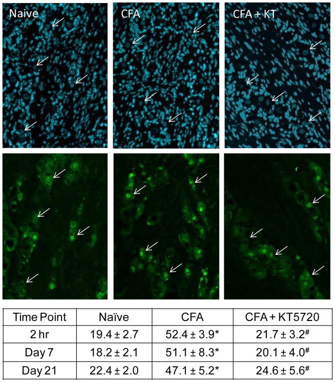
Fig. 5.
Increased nuclear p-ERK expression in trigeminal ganglion neurons in response to CFA was inhibited by KT5720. Representative images of sections from the V3 region of trigeminal ganglia obtained from naïve, CFA, and CFA + KT5720 treated animals at 2 hr, 7 days, and 21 days after injections are shown. All cell nuclei are identified by the nuclear dye DAPI (top panels). The same images were co-stained for p-ERK (bottom panels). Arrows indicate neuronal cell body nuclei identified by DAPI. The average percent ± SEM of p-ERK positive neuronal nuclei, as identified by DAPI staining, for each condition is reported in the table. * P < .05 when compared to naïve levels and #P < .05 when compared to saline control levels.

