
FIGURE 2.
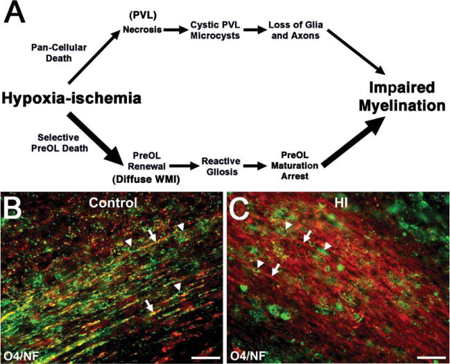
Myelination failure in chronic diffuse white matter injury (WMI) coincides with lesions highly enriched in reactive astrocytes, activated microglia, and oligodendrocyte progenitors (preOLs) that are arrested in their maturation. (A) Distinctly different pathogenetic mechanisms mediate abnormal myelination in necrotic lesions (periventricular leukomalacia [PVL]; upper pathway) versus lesions with diffuse WMI (lower pathway). Hypoxia–ischemia (HI) is illustrated as one potential trigger for WMI. More severe HI triggers white matter necrosis (upper pathway) with pancellular degeneration that depletes the white matter of glia and axons. Severe necrosis results in cystic PVL, whereas milder necrosis results in microcysts. Milder HI (lower pathway) selectively triggers early preOL death. preOLs are rapidly regenerated from a pool of early preOLs that are resistant to HI. Chronic lesions are enriched in reactive glia (astrocytes and microglia/macrophages) generating inhibitory signals that block preOL differentiation to mature myelinating oligodendrocytes. Myelination failure in diffuse WMI thus results from preOL arrest rather than axonal degeneration. The molecular mechanisms that trigger preOL arrest are likely to be multifactorial and related to factors intrinsic and extrinsic to the preOLs. Note that the lower pathway is the dominant one in most contemporary preterm survivors, whereas the minor upper pathway reflects the declining burden of white matter necrosis that has accompanied advances in neonatal intensive care. (B) Typical appearance of normal early myelination of axons in a perinatal rodent at postnatal day 10.80 Axons are visualized by staining for neurofilament protein (NF; red). Early myelination of axons is visualized with the O4 antibody (green). (C) preOL arrest in a chronic white matter lesion where numerous preOLs (green) are seen, but the axons (red) are diffusely unmyelinated. Scale bars = 100μm.