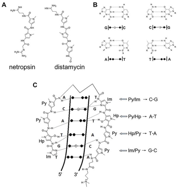
Figure 10.
Design of polyamides to target DNA sequence. (A) Structures of the naturally occurring polyamides netropsin and distamcyin and (B) Watson—Crick hydrogen-bonding patterns in the DNA minor groove. The black circles represent lone electron pairs, and circles containing an H represent the 2-amino group of guanine. R represents the sugar backbone of DNA; (C) binding model between ImHpPyPy-γ-mHpPyPy-β-Dp and a 5′-TGTACA-3′/3′-TGTACA-5′ sequence. Hydrogen bonds are shown as dashed lines.

