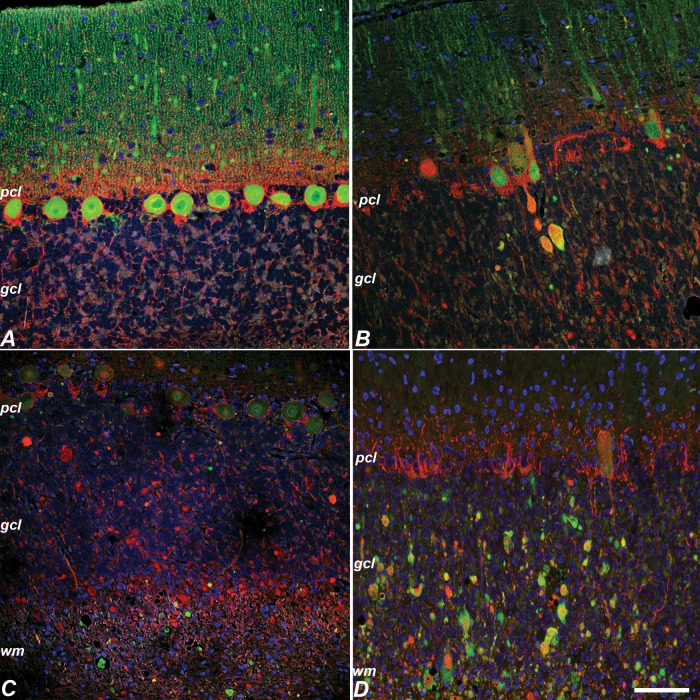
FIGURE 5.
Neurofilament labeling in normal control and NPC1 cat cerebellar cortex. (A) Neurofilament (red in all panels) was not detected in control Purkinje cells (calbindin, green). (B) In NPC1, rare neurofilament and calbindin colabeling was seen in Purkinje axon swellings (calbindin, green). (C, D) However, most axonal swellings were neurofilament-positive and calbindin-negative (calbindin, green) (C). The great majority of neurofilament positive swellings colabeled with LC3 (LC3, green) (D). pcl, Purkinje cell layer; gcl, granular cell layer; wm, cerebellar white matter. Scale bar: 100 µm.

