
Figure 2.
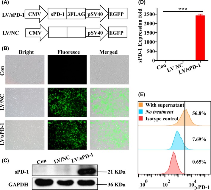
Confirmation of soluble programmed death receptor‐1 (sPD‐1) expression post‐lentivirus infection. A, Schematics of sPD‐1 overexpressing lentivirus (LV/sPD‐1) and negative control lentivirus (LV/NC). EGFP, enhanced green fluorescent protein. B, Verification of infected efficiency of lentivirus based on fluorescence microscopy of EGFP expression on 4T1 cells (magnification ×200). C, Western blotting analysis of sPD‐1 protein expression on 4T1 cells infected by LV/NC and LV/sPD‐1, with 4T1 cells as a control. D, qRT‐PCR analysis of sPD‐1 RNA expression on 4T1 cells infected by LV/NC and LV/sPD‐1, with 4T1 cells as a control. ***P < .001. E, 4T1 cells were pretreated with IFN‐γ to increase programmed death ligand‐1 expression. Culture medium from 4T1/sPD‐1 cells was added to 4T1 cells as mentioned above and incubated for approximately 30 min before flow cytometry assay for detecting ratio of PD‐1+ 4T1 cells. PD‐1, programmed death receptor‐1