
Figure 4.
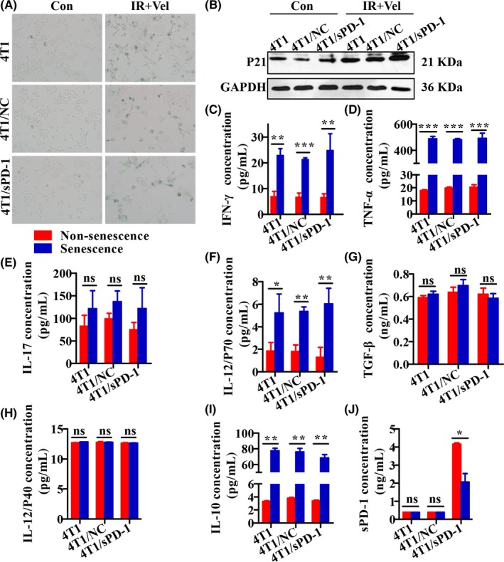
Senescence‐associated secretory phenotype of 4T1, 4T1/NC, and 4T1/sPD‐1 cells treated with radiation and veliparib. NC, negative control; sPD‐1, soluble programmed death receptor‐1. A, SA‐β‐gal staining was used to detect senescence. Bright blue cells were regarded as senescent (magnification ×200). B, Western blotting analysis of senescence marker p21 protein expression. C‐J, Secretion of interferon (IFN)‐γ, tumor necrosis factor‐α (TNF)‐α, interleukin (IL)‐17, IL‐12P70, transforming growth factor‐β (TGF‐β), IL‐12P40, IL‐10 and sPD‐1 was compared between non‐senescent and senescent cells (ns, non‐significant, *P < .05,**P < .01,***P < .001)