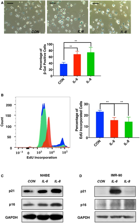
Representative images (above) and quantification (below) of SA‐βGal staining of IL‐6‐ and IL‐8‐treated HBE cells. HBE cells were treated with CD36‐dependent cytokines IL‐6, IL‐8, or PBS at a concentration of 50 ng/ml for 9 days. Cells were then fixed and stained for SA‐βGal activity. Scale bars, 50 μm. Data are reported as the mean ± SEM (n = 3). **P < 0.01, one‐way ANOVA.
Proliferation analysis of IL‐6‐, IL‐8‐, or PBS‐treated HBE cells. HBE cells were cultured with recombinant cytokines as described in (A). Then, cells were further treated with 10 μM EdU for 2 h prior to collection, staining, and analysis by flow cytometry. Data are presented as the mean ± SEM of three biological replicates. P‐values were calculated based on three independent experiments. **P < 0.01, one‐way ANOVA.
Expression of cyclin‐dependent kinase inhibitors p21 and p16 was analyzed using the lysates from samples as described in (A). Blots shown are representative of three independent biological replicates.
IMR90 fibroblasts were treated with 50 ng/ml IL‐6, IL‐8, or PBS for 9 days. Cell lysates were then immunoblotted using antibodies recognizing p16, p21, or GAPDH, as indicated.

