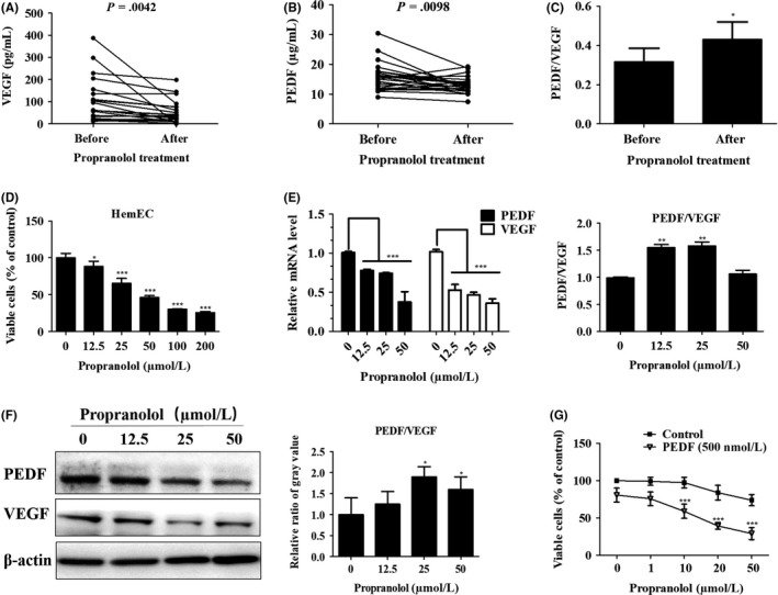
Figure 7.

Pigment epithelium‐derived factor (PEDF) and propranolol combination treatment achieves greater suppression of hemangioma‐derived endothelial cell (HemEC). A,B, Paired blood serum samples of 20 patients before and after 1 mo of oral propranolol treatment. A, Vascular endothelial growth factor (VEGF) and (B) PEDF expression in the serum was determined using human PEDF and VEGF‐A ELISA kits and paired t test analysis was carried out. C, Relative PEDF and VEGF ratio was calculated in absolute terms and analyzed with paired t tests. Results are presented as the mean ± SEM, *P < .05. D, Different concentrations of propranolol were added to HemEC for 24 h, and CCK‐8 assays were carried out to measure cell vitality. *P < .05,***P < .001 vs the 0‐concentration group. E, qRT‐PCR analysis of VEGF and PEDF expression in HemEC after treatment with propranolol at the indicated concentrations for 24 h. Histogram indicates the relative PEDF and VEGF ratio in terms of mRNA levels, and results are shown as the mean ± SD of triplicates.**P < .01, ***P < .001 vs the 0‐concentration group. F, Western blotting analysis of VEGF and PEDF production in HemEC after treatment with propranolol at the indicated concentrations for 48 h. β‐actin, control protein. Histogram indicates the relative ratio of the PEDF and VEGF gray values from the western blots, and the results are shown as the mean ± SD of triplicates. *P < .05 vs the 0‐concentration group. G, PEDF/VEGF ratio was artificially modified with treatments comprising combinations of PEDF (500 nmol/L) and different concentrations of propranolol in HemEC for 48 h, and cell viability was measured with CCK‐8 assays. Results are shown as the mean ± SD. ***P < .001 vs the control group
