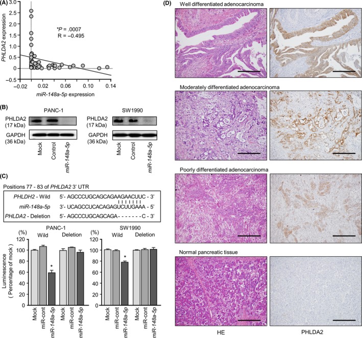
Figure 5.

Direct regulation of PHLDA2 by miR‐148a‐5p in pancreatic ductal adenocarcinoma (PDAC) cell lines and immunohistochemical staining of PHLDA2 protein in PDAC clinical specimens. A, Expression levels of PHLDA2 and miR‐148a‐5p were negatively correlated. B, PHLDA2 protein expression in PDAC cell lines was evaluated by western blot analyses 96 h after transfection with miR‐148a‐5p. GAPDH was used as a loading control. C, miR‐148a‐5p binding sites in the 3′‐UTR of PHLDA2 mRNA. Dual luciferase reporter assays using vectors encoding the putative miR‐148a‐5p (positions 77‐83) target site of the PHLDA2 3′‐UTR for both wild‐type and deleted regions. Normalized data were calculated as ratios of Renilla/firefly luciferase activities, *P < .05. D, Immunohistochemical staining of PHLDA2 in PDAC specimens. All differentiated types of PDAC (well, moderate and poor) showed strong intracellular immunoreactivity (left panel, H&S staining; right panel, PHLDA2 staining; original magnification, ×200; scale bars, 200 μm)
