Figure 2.

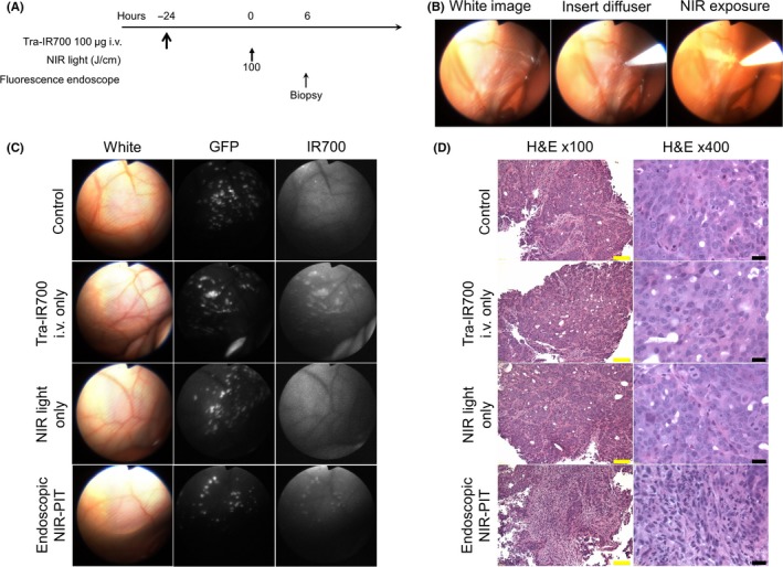
In vivo fluorescence real‐time endoscopic imaging and histological near infrared photoimmunotherapy (NIR‐PIT) effect. A, Treatment regimen is shown. Biopsy specimens were obtained 6 hours after NIR‐PIT. B, In vivo real‐time endoscopic intraperitoneal imaging of N87GFP‐luc tumor‐bearing mice. Peritoneal cavity was exposed to NIR light using an optical diffuser through the endoscope. C, In vivo fluorescence real‐time endoscopic intraperitoneal imaging of N87GFP‐luc tumor‐bearing mice. Tumors demonstrate GFP and IR700 fluorescence in mice administered tra‐IR700. In the absence of tra‐IR700 no IR700 fluorescence was seen. After NIR‐PIT, IR700 fluorescence decreased. D, Biopsy specimens stained with H&E demonstrate few scattered clusters of damaged tumor cells within a background of diffuse cellular necrosis and micro‐hemorrhage with infiltration of inflammatory cells consistent with acute granulation in endoscopic NIR‐PIT group, while no obvious damage was observed in control groups, including tra‐IR700 alone without NIR light and NIR light alone without tra‐IR700 groups. Yellow scale bars = 100 μm. Black scale bars = 20 μm
