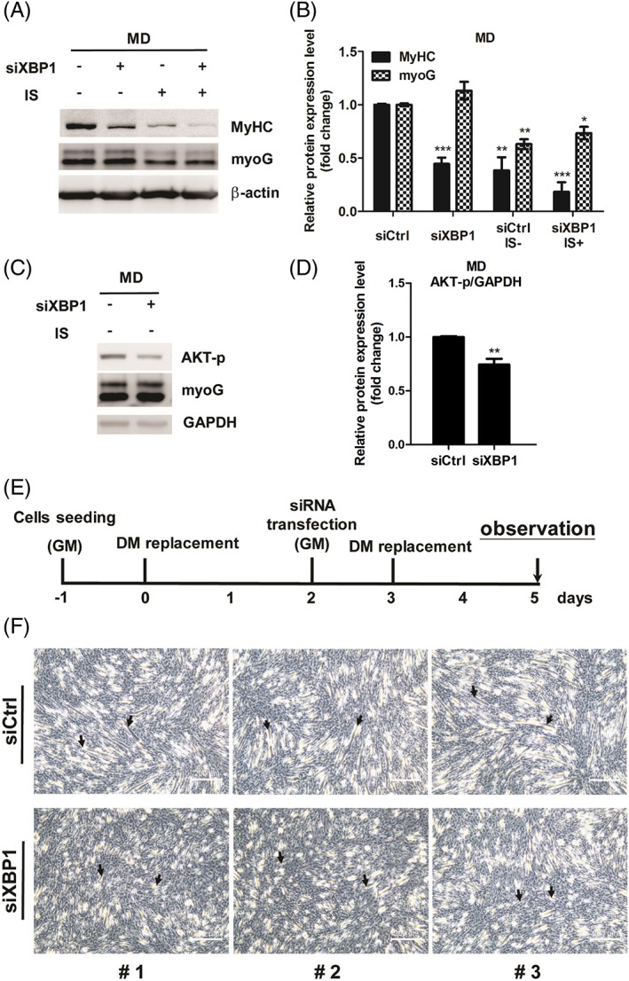
Figure 6.

Promyogenic role of XBP1 in myoblast differentiation (MD). (A–D) The expression of MyHC, myoG, and phospho‐AKT in XBP1‐dificent cells is detected by western bot and quantified. (E) Diagram illustrates timeline of experiment. (F) Phase contrast photomicroscopy revealed differentiated morphology in XBP1‐deficient myoblasts compared with ordinary myoblasts. The arrows indicate the fusion of myoblasts. Random views of three independent experiments at a magnification of ×100 were shown, scale bars = 200 μm.
