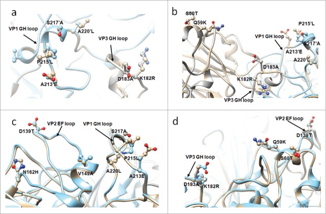
Figure 6.

The adjacent variant sites are displayed in the structure. (a) VP1 GH loop and VP3 GH loop, (b) VP1 GH loop, VP3 GH loop and residue 59, 60 of VP3, (c) VP1 GH loop and VP2 EF loop, (d) VP2 EF loop and residue 59, 60 of VP3. The structure of circulating recombinant strain colored in golden, G10 colored in cyan. 220′ indicates the residue 220 of an adjacent protomer.
