
Figure 6.
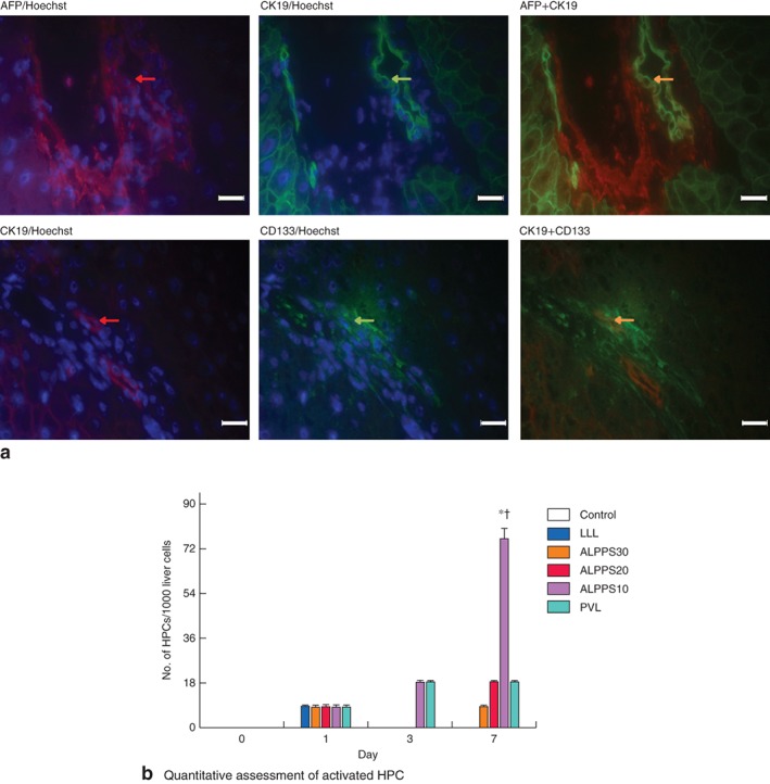
Representative expression of hepatic progenitor cell (HPC) activation in the future liver remnant after surgery. a Double immunofluorescence staining with α‐fetoprotein (AFP) (red) plus cytokeratin (CK) 19 (green) or CK19 (red) plus cluster of differentiation (CD) 133 (green), and Hoechst nuclear staining (blue) (magnification ×400, scale bars 20 μm). Expression of activated HPC is indicated by simultaneous positive staining (yellow) of both CK19 and AFP/CD133 (arrows). b Quantitative assessment of the number of HPCs per 1000 liver cells on days 1, 3 and 7 after step 1 surgery following left lateral lobe (LLL) resection, Associating Liver Partition and Portal vein ligation for Staged hepatectomy (ALPPS) and portal vein ligation (PVL). Values are mean(s.d.) (4–5 animals per group). *P < 0·050, ALPPS10 versus LLL, ALPPS30 and ALPPS20 (ANOVA); †P < 0·050, ALPPS10 versus PVL (Student's t test)