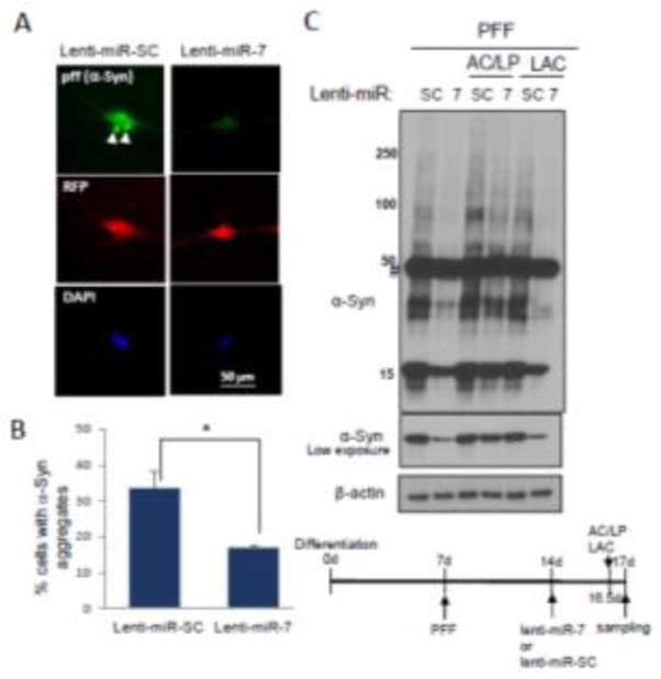
Fig. 4.

miR-7 decreases the levels of intracellular α-Syn transported from the outside as pff. Differentiated ReNcell VM cells were treated with pff (100ng). After 7 days, these cells were transduced with either lenti-miR-SC or lenti-miR-7 for another 3 days. (A) Representative figures showing miR-7-induced decrease of α-Syn aggregates (pff). Lentivirus-infected cells are detected as red fluorescence and α-Syn expression is shown as green fluorescence. α-Syn aggregates are indicated as white arrowheads. (B) Cells having α-Syn aggregate(s) were counted in 7 randomly selected fields comprising 10–20 RFP-positive cells (lentivirus-transduced cells). The data represent means ± SEM. * p < 0.01. (C) During the period of lentiviral transduction, cells were further treated with AC (NH4Cl (10 mM)), LP (leupeptin (100 μM)) or LAC (lactacystin (5 μM)) for 12 h before harvest. Cells were subjected to Western blot analyses to detect α-Syn and β-actin. Time-course showing differentiation, PFF exposure, viral transductions and chemical treatments is presented. These results are representative of three separate experiments.
