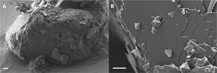
Fig. 1. Secondary electron mapping (SEM) images of a loose diamond extracted from the run products of sample H3908 by mechanical deposition on a stub covered with carbon tape before FIB preparation.

(A) Diamond growth covers the whole initial seed. (B) Close-up of the surface exhibiting the formation and trapping of new spontaneously nucleated diamond crystals as inclusions. Scale bar, 2 μm.
