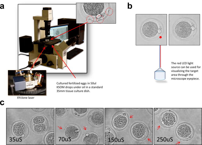
Fig. 1. An Infrared Laser Perforation System to Perforate the Zona.
(a) The laser is housed in a lens and can be attached to the turret of most inverted microscopes. The laser permits perforation of targeted areas in zona without micromanipulation of the embryos. Perforations in the zona are circled in red. Perforation size is controlled by the duration of laser firing, typically 250 microseconds. Photographs provided by Steven R. McCaw, NIEHS Multimedia Services. (b) The factory-aligned laser is locked in place and contains a red LED light source for visualizing the target area. The controlling software allows for rapid laser fire while looking through the microscope eyepieces. The fertilized eggs are laser treated while free floating in KSOM drops – before (left) and after (right) images of an embryo. (c) Size of the perforation by the XYclone laser directly affected the efficiency of gene delivery. Perforations generated by the laser treatments ranged from 35uS to 250uS. Larger perforations allowed for a more effective viral transduction. The results of seven independent experiments are shown in Table 2 summarizing the number of blastocysts and their fluorescence used in the experiments.

