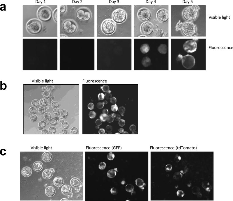
Fig. 2. En Masse Gene delivery to Fertilized Eggs.
(a) Development of C57BL/6J embryos from harvest (Day1) until the time of blastocyst implantation (Day 5). The embryos were perforated by the Xyclone laser (3 holes, 250uS) and treated with 2uL of high titer lentivirus (108–109 TU/ml) in 50uL KSOM drops. The presence of fluorescence was detected in days 3 and 4 and persisted until the time of implantation. The data is representative of five independent experiments - 49 blastocysts formed out of 82 treated fertilized eggs, 44 out of 49 blastocysts expressed GFP. (b) Most blastocysts infected with the lentivirus delivering GFP expressed fluoresce at the time of implementation. Cultured blastocysts from Crl:CD-1 (ICR) strain of mice are shown in the figure. (c) Co-infection of fertilized eggs with viruses expressing either GFP or tdTomato readily expressed both genes after 4 days. The pattern of expression for GFP and tdTomato in infected blastocysts are not identical although both genes are expressed from the EF1α promoter.

