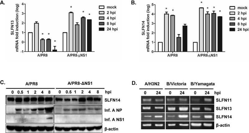
Fig. 3.
Induction of SLFN13 and SLFN14 following influenza virus infection. A549 cells were infected with the human influenza virus strain PR8 or PR8/delNS1 (multiplicity of infection = 1) for the indicated lengths of time. Host mRNA expression was measured by real-time qRT-PCR for SLFN13 (A) and SLFN14 (B). The expression of target genes was normalized to that of GAPDH. Expression in the mock-infected control was set to 1, and other samples were normalized to this value. Data are shown as the mean ± SEM of three independent experiments. Statistical analysis: *p < 0.05 compared with mock-infected cells at each timepoint. (C) SLFN14 protein levels were measured in influenza A virus-infected mouse macrophages RAW 264.7 cells. The images shown are representative of three independent experiments. (D) A549 cells were infected with clinical influenza virus strains, including seasonal A/H3N2, B/Victoria, and B/Yamagata, at a multiplicity of infection of 0.1 for 24 h, and RT-PCR was performed.

