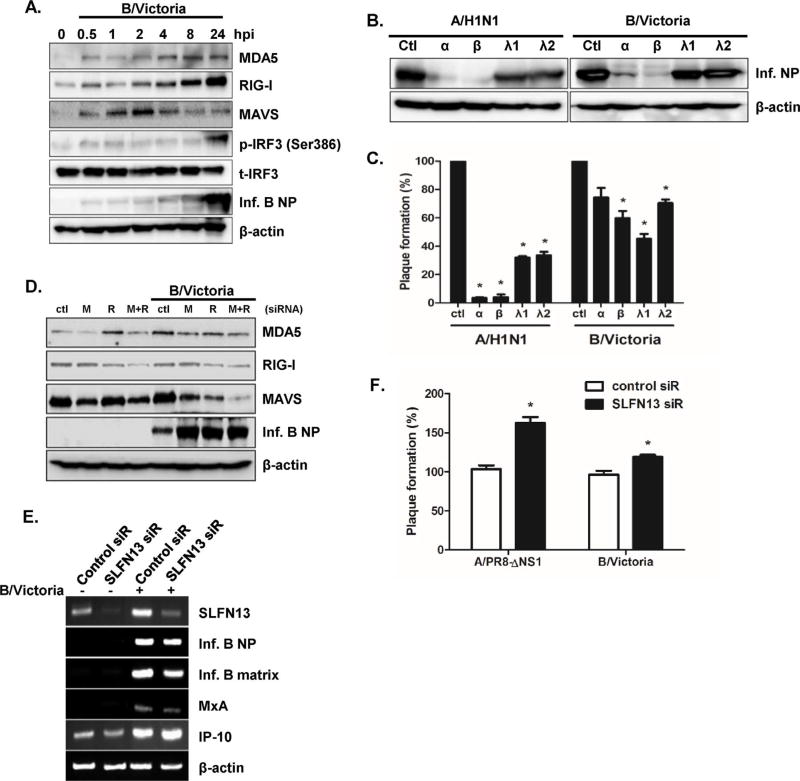
Fig. 7.
SLFN13 knockdown results in increased viral replication in response to influenza B virus infection (A) A549 cells were infected with human influenza B/Victoria virus (MOI = 0.1) for 24 h. Protein levels of RIG-I, MDA5, MAVS, phospho-IRF3, and viral NP were analyzed by western blotting. The images shown are representative of three independent experiments. (B, C) Recombinant human IFN-α (10 ng/mL), IFN-β (10 ng/mL), IFN-λ1 (10 ng/mL), and IFN-λ2 (10 ng/mL) were added to cells. The next day, cells were infected with A/H1N1 or B/Victoria virus, and viral NP expression and viral titers were measured. Progeny viral titers were calculated and expressed as plaque-forming units (PFU)/mL. Control-treated cells were normalized to 100%, and data are presented as the percentage relative to the control treated samples. Data are shown as means ± SEM of two independent experiments. (D) A549 cells were transfected with control or RIG-I (R) or MDA5 (M)-specific siRNA, and the knockdown efficiency was measured by western blotting. Levels of viral NP proteins were measured, and anti-actin monoclonal antibody was used as a loading control. The images shown are representative of three independent experiments. (E) Knockdown of SLFN13 was performed with SLFN13-specific siRNA transfection. mRNA expression of SLFN13, MxA, IP-10, influenza B NP, and influenza B matrix genes was measured by RT-PCR. (F) Progeny viral titers were calculated and expressed as plaque-forming units (PFU)/mL. Data are shown as means ± SEM of three different experiments and are presented as the percentage relative to the control siRNA sample. Statistical analysis: *p < 0.05 compared with control siRNA-transfected cells.

