
Figure 7.
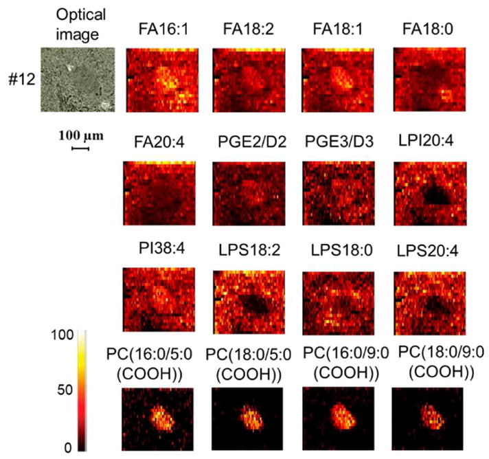
Negative mode nano-DESI imaging of a small region (500*420 Gm2) of mouse pancreatic tissue. All the images were normalized to the internal standard (D8-arachidonic acid).
Official websites use .gov
A
.gov website belongs to an official
government organization in the United States.
Secure .gov websites use HTTPS
A lock (
) or https:// means you've safely
connected to the .gov website. Share sensitive
information only on official, secure websites.
Negative mode nano-DESI imaging of a small region (500*420 Gm2) of mouse pancreatic tissue. All the images were normalized to the internal standard (D8-arachidonic acid).