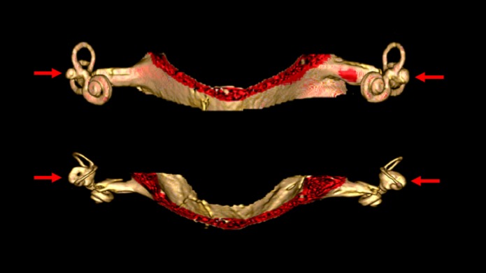
Figure 4.
High resolution three-dimensional reconstruction MRI of the internal auditory canals and inner ear structures of a patient with bilateral vestibular weakness from bilateral labyrinthine dysplasia. The top image is in the coronal aspect. The bottom image is in the axial aspect. In these images it is evident that the horizontal canals are dysplastic, with the horizontal canal and vestibule appearing as a single abnormal structure on each side (indicated by the arrows). The superior and inferior canals are present as true canals, but are somewhat hypoplastic.

