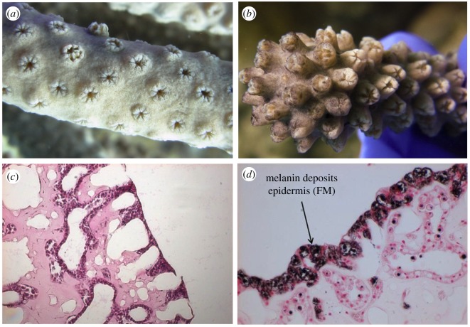
Figure 1.
Macro and histological views of healthy (a,c) and diseased (b,d) Eunicea. (a) Close up of a healthy colony of Eunicea lacking any characteristic black pigmentation associated with the disease. (b) Close up of a colony of Eunicea infected with EBD; note the characteristic black pigmentation of the tissue. (c) Standard haematoxylin and eosin stained histology slides of apparently healthy sample showing good tissue organization and normal staining characteristics (pink staining) and (d) slides of tissue from an infected colony stained with Fontana-Masson showing the epidermis with melanin deposition.

