
Figure 1.
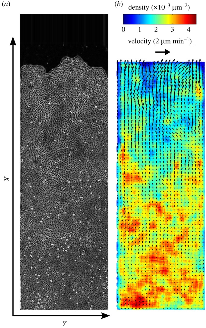
Cell migration. (a) A monolayer of MDCK cells, initially confined, is released. It expands (electronic supplementary material, movies S1–S3) along the adhesive strip towards empty space (direction of increasing x). Mitomycin C is added to inhibit divisions. Phase-contrast image of cell contours, taken at t=11 h 30 min (i.e. after approx. 16 h 30 min of migration). Strip total length 4 mm (most of it is visible here), width 1 mm. (b) Corresponding 2D fields of cell velocity and density. Scale arrow: .