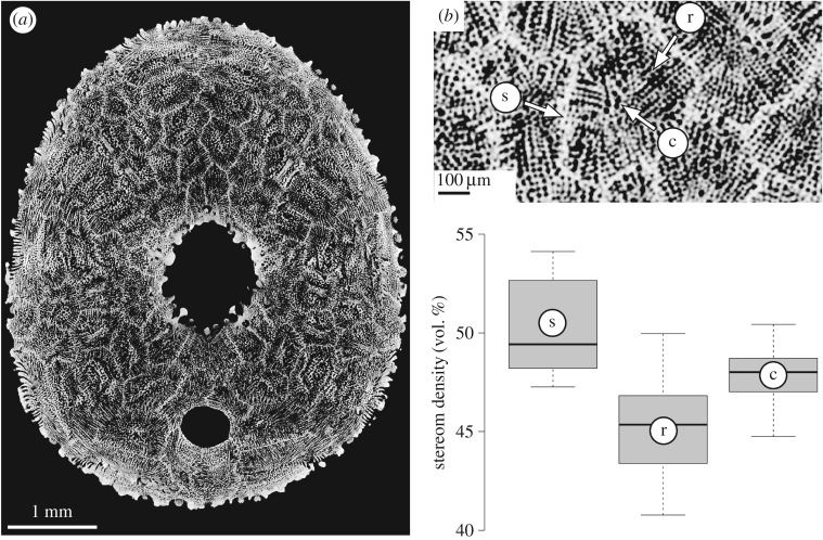
Figure 4.
Stereom differentiation in E. pusillus [GPIT/EC/00740:gg-al-1.73]. (a) Micro-CT section of the oral side showing the mosaic of plates. (b) Close-up indicating three analysed regions of the plate: c, unordered labyrinthic stereom at the plate's centre; r, directional galleried stereom at the plate's rim; s, directional galleried stereom within sutures. (c) Comparison between three prominent areas of the plates.

