
Figure 5.
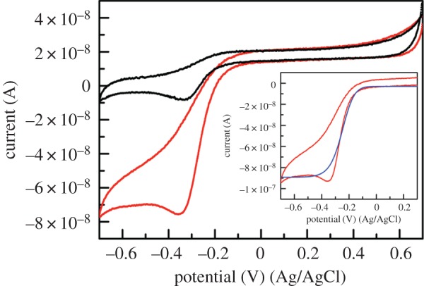
Representative CVs for a NAcMP–HXT–Au electrode in 0.1 M phosphate buffer (pH 7) (black lines) or in the presence of 0.33 µM H2O2 (red lines). Scan rate 10 mV s−1, temperature 22 ± 2°C. The inset compares the background-subtracted experimental CV (red lines) and the theoretical current–voltage curve during the forward scan (blue line). Owing to a quasi-reversible direct electron transfer reaction, replication of the reversed-scan wave was unsatisfactory.