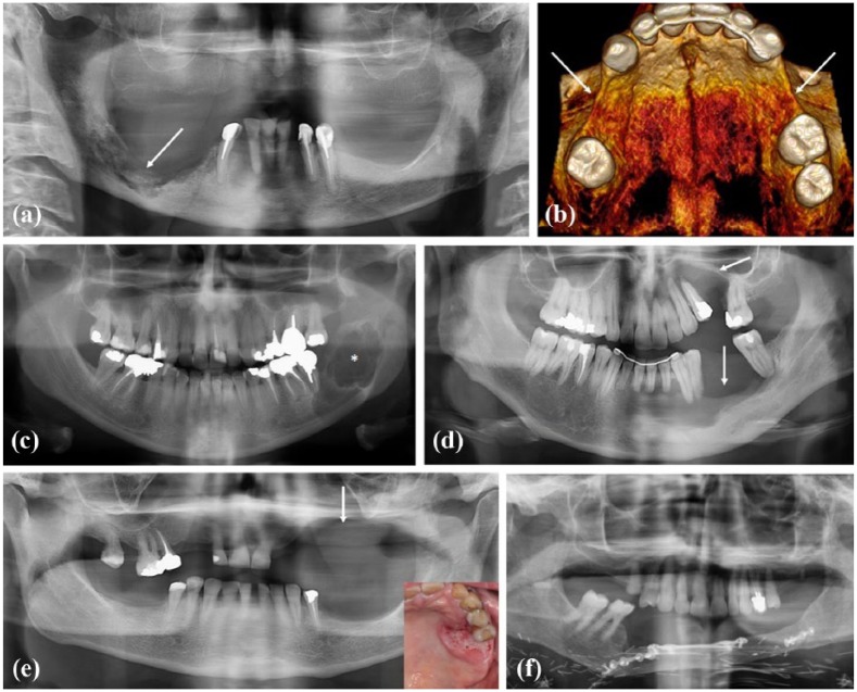
Figure 1.
Various origins of bone defects: (a) Panoramic X-ray. Bone defect of the mandible right body corresponding to an osteonecrosis of the jaw in relation to denosumab taking. (b) 3D-reconstructed view of upper jaw. Bilateral bone defect of premolar regions associated to tooth agenesis in a young adult presenting a WNT10A gene mutation. (c) Panoramic X-ray. Bone defect (radiolucency, *) of the mandible right ramus corresponding to an ameloblastoma, an odontogenic aggressive benign tumor. (d) Panoramic X-ray. Bone defect (radiolucency, arrows) of upper and lower jaws corresponding to a trauma. (e) Panoramic X-ray. Bone defect (arrow) of upper jaw after resection surgery of a gingival squamous cell carcinoma (clinical view, left corner). (f) Reconstruction of the mandible by autogenous bone (fibula) following an invasive squamous cell carcinoma of the gingiva.

