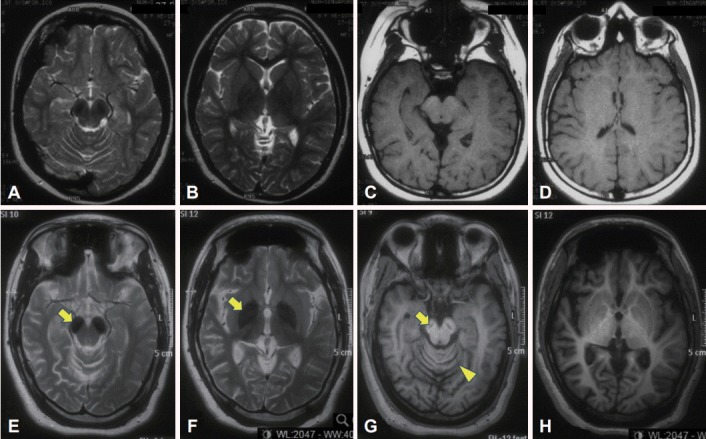
Figure 1.

Brain MRI scans performed at ages eight (top panel) and 24 (bottom panel). The first MRI was initially reported as normal but in retrospect shows bilateral T2 hypointensity in the substantia nigra (A). However, the first MRI shows normal signal in the globus pallidus (B) (more prominent nigral vs. pallidal involvement is typical of beta-propeller protein-associated neurodegeneration) [1]. There is also mild thinning of the posterior corpus callosum, seen on the sagittal T1 image (not shown). Later, marked T2 hypointensity in the substantia nigra and cerebral peduncle (arrow) (E) and globus pallidus (arrow) (F) has developed bilaterally. There is a “slit” of T1 hypointensity in the nigra with a faint rim of hyperintensity, the so-called “halo” sign (arrow) (G), as well as global cerebral atrophy (G and H, compared to C and D) and cerebellar atrophy (arrowhead) (G).
