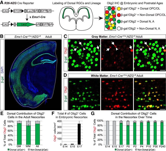
Figure 1.
Most Olig2+ cells in the neocortex are dorsally derived beginning at embryonic ages. A, Schematic of the genetic lineage-tracing approach to distinguish dorsally derived cells in the neocortex from ventrally derived cells. Emx1-Cre mice were crossed to R26-NZG reporter mice to permanently label all Emx1+ progenitors and their offspring (β-gal+) and identify oligodendrocyte lineage cells by immunostaining for Olig2. OPC, oligodendrocyte precursor cell; OL, Oligodendrocyte; N, neuron; A, astrocyte. B, Representative image of β-gal+ cells in the adult forebrain, showing the Emx1-Cre recombination pattern in dorsally derived, but not ventrally derived, structures. C, Examples of Olig2+ (red) cells in the gray matter of the adult somatosensory cortex. Arrows denote dorsally derived (β-gal+) Olig2+ cells and the arrowhead denotes a ventrally derived (β-gal−) Olig2+ cell. D, Examples of Olig2+ (red) cells in the white matter of the somatosensory cortex that are β-gal+ (green) and therefore dorsally derived. E, Graph of the average (±SEM among biological replicates) percentage of Olig2+ cells in the adult neocortex that were dorsally derived (β-gal+) versus not dorsally derived (β-gal−). Quantification was performed in the somatosensory cortex, separately in the white matter (WM), gray matter (GM), or combined (All). F, Quantification (average ± SEM among biological replicates) of total number of Olig2+ cells in the somatosensory cortex (white and gray matter combined) at early embryonic ages. Total numbers of Olig2+ cells were counted per mm2. G, Graph of the average (±SEM among biological replicates) percentage of Olig2+ cells in the somatosensory cortex (white matter and gray matter combined) that were dorsally derived (β-gal+) versus not dorsally derived (β-gal−) at different time points in embryonic (E) and postnatal (P) development. Scale bar, 20 μm.

