Figure 5.

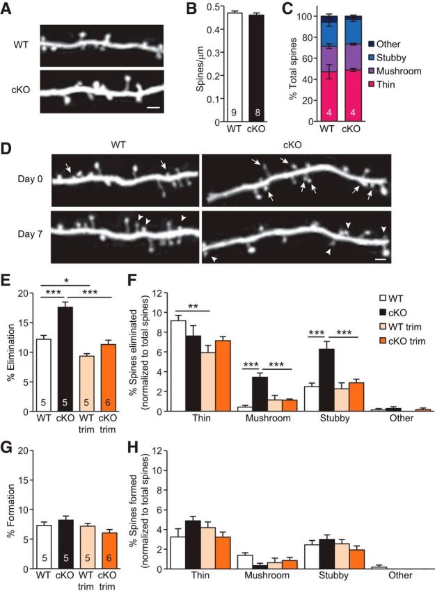
Experience-dependent elimination of spines is elevated in L5 PNs of P30 CaMKIIα-RARα cKO mice. A, Representative images taken by in vivo transcranial imaging of YFP in WT and CaMKIIα-RARα cKO mice. Scale bar, 2 μm. B, Quantification of spine density in P30 WT and CaMKIIα-RARα cKO mice (7–12 dendrites per animal were analyzed, Student's t test). C, Distribution of spine types in WT and CaMKIIα-RARα cKO mice (two-way ANOVA). D, Repeated imaging of the same dendritic branches over a 7 d interval in the barrel cortex of WT and cKO mice under control conditions reveal newly formed spines (arrowheads), and eliminated spines (arrows). Scale bar, 2 μm. E, Quantification of the percentages of spines eliminated over 7 d in the barrel cortex of WT and cKO mice under control and trimmed conditions (Trim; *p < 0.05, ***p < 0.001, two-way ANOVA). F, Quantification of the percentages of eliminated spine types normalized to total spines eliminated (**p < 0.01, ***p < 0.001, two-way ANOVA). G, Quantification of the percentages of spines formed over 7 d in the barrel cortex of WT and cKO mice under control and whisker-trimmed conditions (two-way ANOVA). H, Quantification of the percentages of formed spine types normalized to total spines formed (two-way ANOVA). In all graphs, data represent average mean ± SEM; n indicates the numbers of mice analyzed.
