Figure 1.

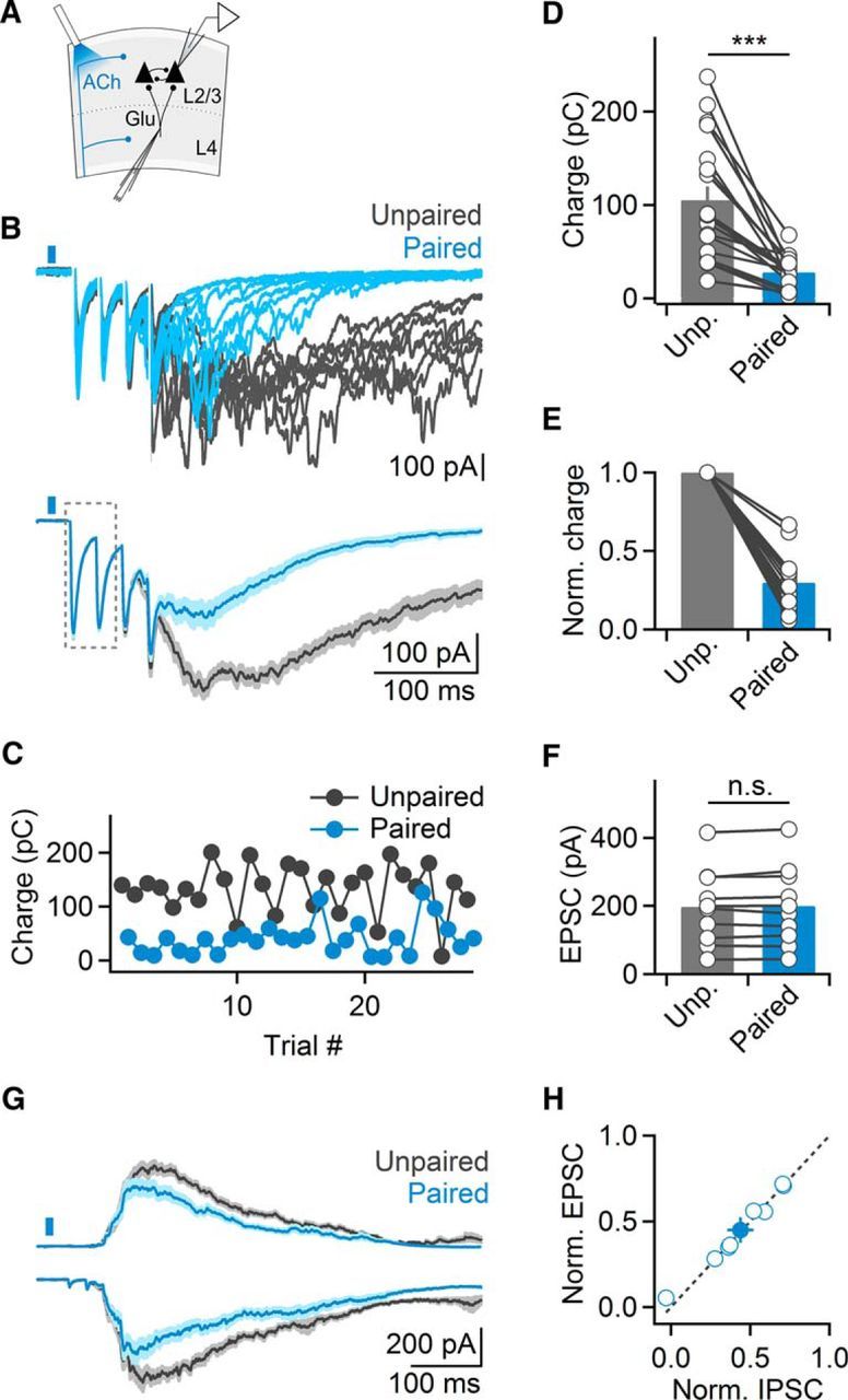
ACh release evoked by single light pulses suppresses evoked cortical recurrent activity. A, Schematic of experimental setup. Cortical recurrent activity was evoked using brief bursts of extracellular stimuli applied in layer 4 and was recorded in layer 2/3 neurons in voltage-clamp. Cholinergic afferents were activated using single light pulses (5 ms), 15 ms before electrical stimulation. B, Top, Representative recording showing multiple trials of recurrent activity, in the absence of (unpaired, black traces) or following optical stimulation (paired, blue traces). Bottom, EPSCs averaged across all unpaired and paired trials. Note lack of amplitude reduction of monosynaptic EPSCs (outlined). C, For the same cell shown in B, plot depicts recurrent activity (quantified as EPSC charge transfer), in paired trials (blue) alternated with unpaired trials (black). D, Summary data showing light-evoked suppression of recurrent activity in layer 2/3 neurons (n = 19 cells). ***p < 0.001. E, Same data as in D, normalized to unpaired responses. F, Summary data showing average amplitude of monosynaptic EPSC evoked by the first two stimuli (n = 10 cells) for unpaired and paired trials. G, Recurrent activity recorded as EPSCs and IPSCs (black, unpaired; blue, paired) from pairs of neighboring layer 2/3 cells, held at −70 and 0 mV, respectively. H, Summary data plotting normalized suppression of EPSCs and IPSCs, for all cell pairs (n = 8). Shaded areas and error bars denote SEM.
