Figure 4.

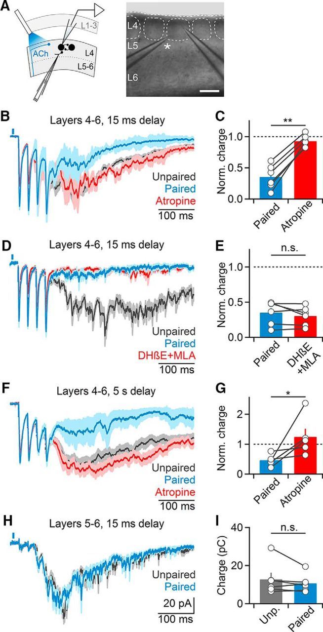
Cholinergic suppression of recurrent activity is layer-specific. A–G, Recordings were performed in slices following surgical removal of layers 1–3. A, Left, Schematic indicating recording arrangement. Right, Bright-field image of slice preparation. Asterisk denotes stimulating electrode. Scale bar, 150 μm. B, Representative recording of layer 4 neuron showing that cholinergic suppression of recurrent activity is entirely mAChR-mediated. C, Summary data (n = 6 cells) showing complete reversal of cholinergic suppression following atropine application. **p < 0.01. D, Representative recording showing that nAChR antagonist application no longer reduces cholinergic suppression (n = 4 cells, DHβE alone: n = 2, DHβE+MLA: n = 2). E, Summary data (n = 5 cells) for experiments as shown in D. F, Cholinergic suppression of recurrent activity is maintained for long delays (5 s) between optical and electrical stimuli and mediated by mAChRs. G, Summary data (n = 5 cells) for experiments as shown in F. *p = 0.02. H, I, Surgical removal of layers 1–4 eliminates cholinergic suppression. Recordings were performed from layer 5 neurons and activity was evoked in the white matter below the same column. H, Representative recording showing EPSCs averaged across paired and unpaired trials. I, Summary data (n = 6 cells) for recordings as shown in H. All shaded areas and error bars denote SEM.
