
Figure 2.
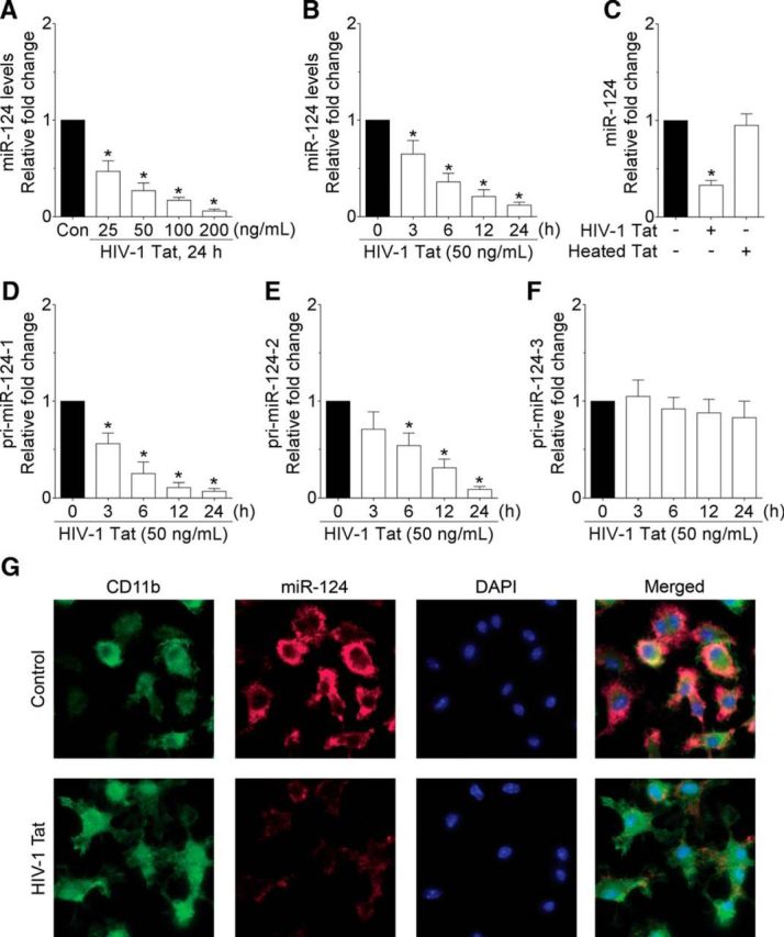
HIV-1 Tat downregulates miR-124 in mouse primary microglial cells. qPCR analysis showing the dose-dependent (A) and time-dependent (B) downregulation of miR-124 expression in HIV-1 Tat exposed to mouse primary microglial cells. C, qPCR analysis showing no change on miR-124 expression in mouse primary microglial cells exposed to heat-inactivated HIV-1 Tat (50 ng/ml) for 24 h. qPCR analysis showing the time-dependent changes on the primary miR-124-1 (D), primary miR-124-2 (E), and primary miR-124-3 (F) in HIV-1 Tat (50 ng/ml) exposed to mouse primary microglial cells. Data are mean ± SEM from six independent experiments. Nonparametric Kruskal–Wallis one-way ANOVA followed by Dunn's post hoc test was used to determine the statistical significance between multiple groups. *p < 0.05 versus control. G, ISH demonstrating decreased expression of miR-124 in HIV-1 Tat (50 ng/ml) exposed mouse primary microglial cells. −, Vehicle treatment (i.e., 1 μl 1 × PBS/ml of medium).