
Fig. 4.
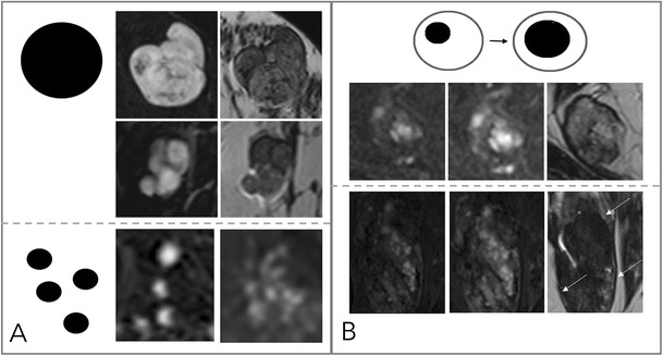
Diagnostic criteria: non-suspicious internal enhancement patterns. A: Homogeneous enhancement suggests benign lesions. Note that even a homogeneous enhancing lesion may show areas of lesser or absent enhancement due to septae and fibrotic parts [A, upper two rows, each early enhanced subtractions (left) and T2w (right)]. Homogeneity is more difficult to assess in non-mass lesions (A, lower row) and includes homogeneous internal morphology. Therefore, this feature was referred to as “stippled” enhancement in the initial BI-RADS lexicon. B: A central or centrifugal enhancement is highly suggestive of a benign lesion. To assess this feature, pre-contrast images need to be considered. While this feature usually applies to mass lesions only (A, upper row, from right to left, early, delayed enhanced, and T2w images), non-mass lesions may also present with this typical benign feature (B, lower row, note the by far larger lesion correlate on the right T2w image, as marked by arrows)