
Fig. 3.
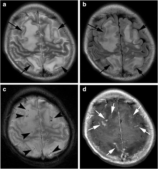
Haemophagocytic lymphohistiocytosis. A 14-year-old girl with myelodysplastic syndrome presented with seizure. a A T2-weighted image shows patchy hyperintensities with swelling in the frontal and parietal lobes (arrows). b FLAIR image shows hyperintensity of these lesions (arrows). c T2*-weighted image shows a number of microhaemorrhages in these lesions (arrowheads). d Post-contrast image shows nodular enhancement along the leptmeninx (white arrows)