
Fig. 4.
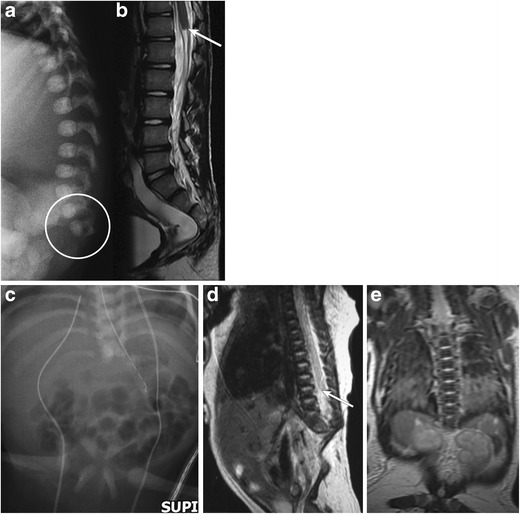
a-b: Caudal/sacral agenesis: Lateral radiograph of the spine (a) in one infant reveals absence of the distal scacrococcygeal segments. Accompanying sagittal T2-weighted MR image (b) of the lumbar spine reveals abnormal truncation of the conus medullaris (arrow). c-e: Caudal agenesis (another neonate): Frontal radiograph of the abdomen (c) in a neonate reveals absence of the entire lumbar and sacrococcygeal spine; the iliac bones are dysmorphic and abnormally close, though separate. Accompanying sagittal (d) and coronal (e) MR images of the spine reveal absence of the lumbar-sacrococcygeal segments of the spine. Abnormal high termination and truncation of the spinal cord is noted (arrow on d)