
Figure 3.
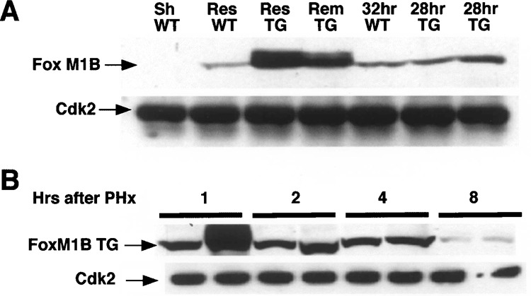
Nuclear FoxM1B transgene protein levels are increased in regenerating liver. Nuclear extracts were prepared from regenerating liver tissue 15 min after PHx, sham-operated (Sh), or regenerating wild-type (WT) and transgenic (TG) liver at the indicated times following PHx, as described previously (33). Lanes marked Res (resected) were nuclear extracts prepared from the liver tissue removed during partial hepatectomy (PHx). Lanes marked Rem (remnant) were nuclear extracts prepared immediately after PHx from the remaining liver tissue. Nuclear protein extracts from regenerating WT liver at 32 h following PHx and duplicate regenerating TG liver at 28 h after PHx were included for comparison. Liver nuclear extract (200 μg) was analyzed by Western blot analysis with polyclonal antibodies against either FoxM1B or Cdk2 (loading control). (A) FoxM1B Western blot analysis of regenerating liver nuclear extracts from WT and TG mice immediately after PHx. (B) Western blot analysis of nuclear extracts prepared from regenerating TG liver prepared from two distinct mice (1, 2, 4, or 8 h following PHx) with FoxM1B antibody.