
Figure 6.
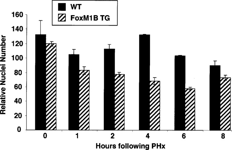
Quantitation of the increase in regenerating transgenic hepatocyte size. We counted and calculated the mean number of hepatocyte nuclei (±SD) per 200× in hematoxylin and eosin (H&E)-stained regenerating WT and TG liver section fields (five separate 200× fields from two distinct regenerating livers). Note that increase in regenerating hepatocyte size is indicated by a decrease in the number of hepatocyte nuclei per 200× field.