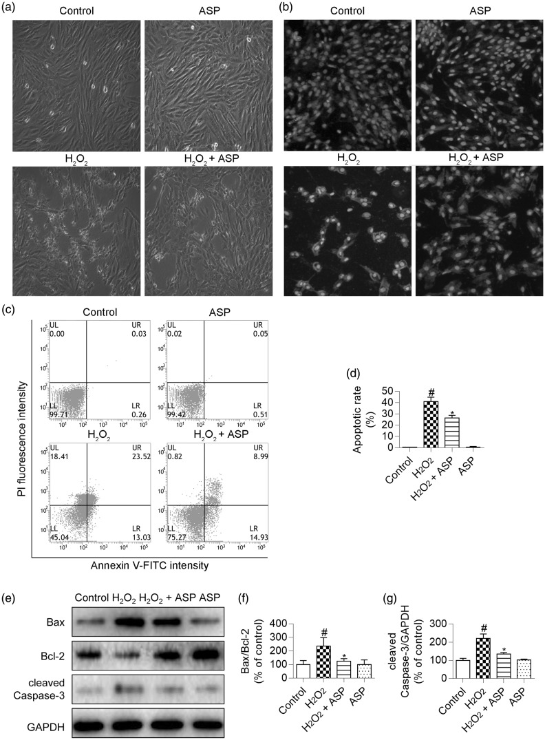
Figure 3.
ASP prevents H2O2-induced apoptosis of H9c2 cells. H9c2 cells were pretreated with 50 µg/mL ASP for 4 hours, followed by incubation with 300 µM H2O2 for 6 hours. (a) Cellular morphology was observed using an inverted microscope. (b) Fluorescence photomicrographs of H9c2 cells stained with Hoechst 33342. (c and d) Apoptosis was assessed by performing flow cytometric analysis after annexin V-FITC/PI double staining, and the number of annexin V-positive cells was quantified. (e) Western blot images of cleaved Caspase-3, Bax, and Bcl-2. (f and g) Densitometry analysis of cleaved Caspase-3 expression and the Bcl-2/Bax ratio. GAPDH was used for normalization. Data are expressed as mean±SD (n = 3). #P <0.05 versus control; *P <0.05 versus H2O2 alone. ASP, Angelica sinensis polysaccharide; H2O2, hydrogen peroxide; fluorescein isothiocyanate/propidium iodide (FITC/PI), GAPDH, glyceraldehyde-3-phosphate dehydrogenase.

