Figure 6.

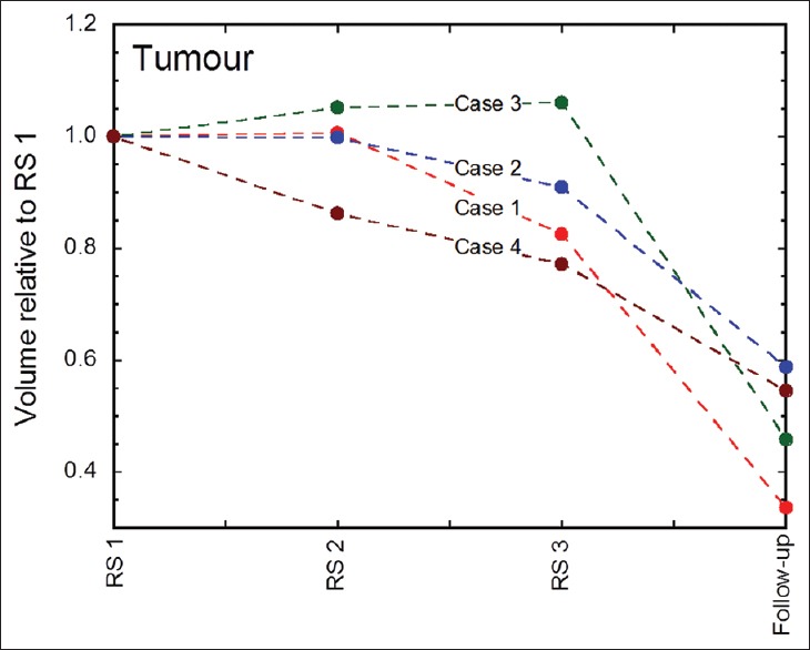
Tumor volume dynamics at each fraction and at 1st follow-up MRI (usually 4 weeks after GKRS 3) for all patients. Case 1 = red line, Case 2 = blue line, Case 3 = green line, and Case 4 = brown line

Tumor volume dynamics at each fraction and at 1st follow-up MRI (usually 4 weeks after GKRS 3) for all patients. Case 1 = red line, Case 2 = blue line, Case 3 = green line, and Case 4 = brown line