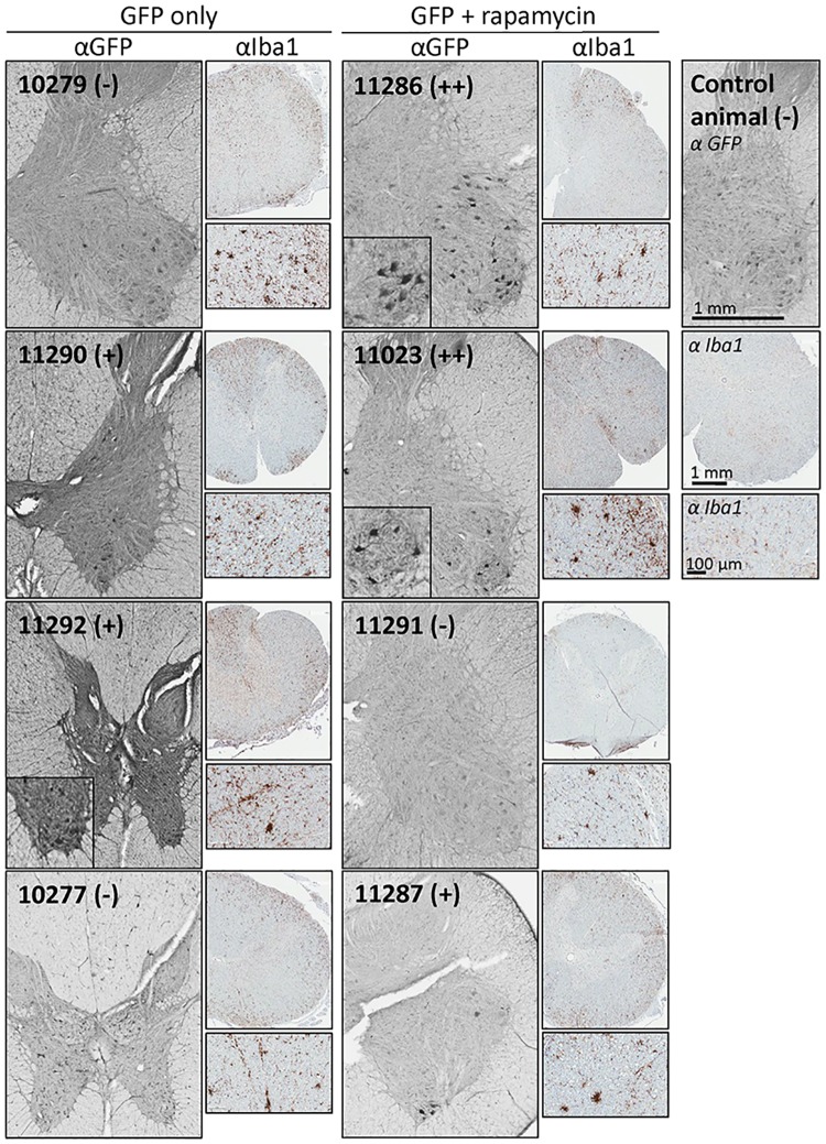
Fig 6. IHC was carried out to visualize GFP expression in the lumbar spinal cord.
Shown are representative 40 micron lumbar spinal cord sections of all study macaques, stained for GFP. Magnified insets show examples of GFP-positive motor neurons. Macaque ID numbers are provided in each panel, along with a qualitative scoring of ventral horn expression. The (-) indicates no GFP-positive staining above that seen in uninjected NHPs; (+) indicates some positive cells observed, above that seen in uninjected macaques; (++) indicates relatively strong expression in a large number of neurons.

